When a doctor suspects heart trouble, the first question is often not "what is wrong?" but "how do we see it clearly?" Two powerful tools dominate this space: Echocardiography, commonly known as an echo, and Cardiac MRI, or CMR. Both look inside your chest without cutting you open, but they work in completely different ways. One uses sound waves; the other uses strong magnets. Understanding the difference between them can help you navigate your diagnosis with confidence and avoid unnecessary delays.
The Basics: Sound Waves Versus Magnets
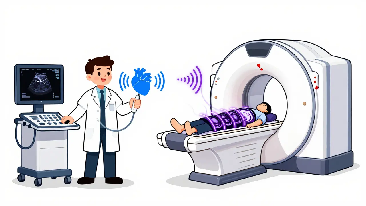
Echocardiography is a non-invasive ultrasound test that creates images of your heart using high-frequency sound waves. Think of it like a sonar used by submarines. A technician places a probe on your chest, sends out sound pulses, and listens for the echoes bouncing back from your heart walls and valves. Because it relies on sound, it works in real-time. You can watch your heart beat on the screen as it happens. It has been around since the 1950s and remains the most common heart imaging test performed worldwide.
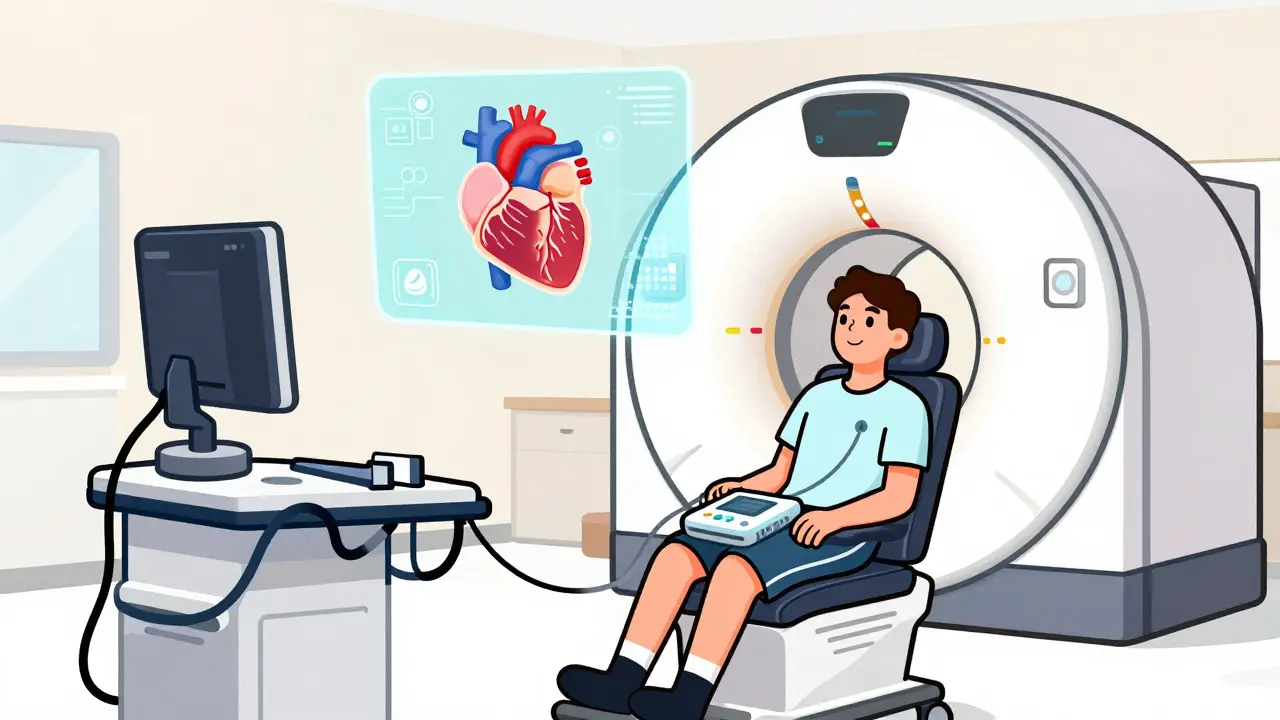
Cardiac MRI is an advanced imaging technique that uses powerful magnetic fields and radio waves to produce detailed cross-sectional images of the heart. This technology emerged in the 1980s and became clinically viable in the 1990s. Instead of sound, it aligns hydrogen atoms in your body using a massive magnet-typically 1.5 to 3.0 Tesla-and then uses radio waves to create highly detailed pictures. Unlike an echo, which gives you a live video, an MRI builds up a static image slice by slice. It takes longer, but the detail is far superior.
| Feature | Echocardiography | Cardiac MRI |
|---|---|---|
| Technology | Ultrasound (Sound Waves) | Magnetic Fields & Radio Waves |
| Speed | Real-time (minutes) | Delayed (30-60 minutes) |
| Cost | $500 - $1,500 | $1,500 - $3,500 |
| Best For | Initial screening, valve function, emergency care | Tissue characterization, precise volume measurement, fibrosis detection |
| Accessibility | Widely available (bedside possible) | Limited to specialized centers |
Why Doctors Start With an Echo
If you have chest pain or shortness of breath, your doctor will almost certainly order an echocardiogram first. Why? Because it is fast, cheap, and safe. According to the American Heart Association, about 15 million echos are performed annually in the United States alone. That is more than ten times the number of cardiac MRIs.
Echos are incredibly versatile. They can be done right at the bedside in an emergency room. If someone collapses with suspected acute aortic dissection, a quick bedside echo can confirm the diagnosis while doctors prepare for surgery. An MRI would take too long and require moving the patient to a specialized scanner, which could be fatal.
The technology also excels at looking at blood flow. Using Doppler techniques, an echo can show if blood is leaking through a valve or if pressure is too high in the lungs. It measures standard metrics like the left ventricular ejection fraction (LVEF)-the percentage of blood pumped out of the main heart chamber-with normal values ranging from 50% to 75%. For routine checks, an echo is usually all you need.
When MRI Becomes the Gold Standard
Sometimes, however, the picture from an echo isn’t clear enough. This happens when patients have "poor acoustic windows." If you are obese, have chronic lung disease, or have thick chest muscles, sound waves struggle to penetrate. The resulting images are fuzzy, making accurate measurements difficult.
This is where Cardiac MRI shines. It does not rely on sound waves traveling through tissue, so body habitus matters much less. More importantly, MRI provides the gold standard for measuring heart volumes and mass. Studies show that echocardiography often underestimates heart size because it relies on geometric assumptions-assuming the heart is a perfect shape. MRI captures the actual 3D volume without those assumptions.
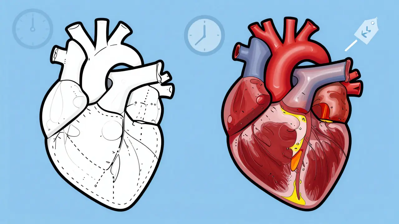
A critical advantage of MRI is its ability to see the heart muscle itself, not just its movement. Using a contrast agent called gadolinium, MRI can detect late gadolinium enhancement (LGE). This highlights scar tissue or fibrosis. If you have had a heart attack, an MRI can show exactly how much muscle died. If you have a condition like hypertrophic cardiomyopathy or cardiac sarcoidosis, MRI can spot inflammation and scarring long before the heart’s pumping function declines. Dr. Susan Joseph of Johns Hopkins University notes that this early detection is critical for intervening before permanent damage occurs.
Accuracy Discrepancies: The Numbers Game
You might wonder why doctors don’t just use MRI for everyone if it is more accurate. The answer lies in the specific numbers. Research published in the Journal of Cardiovascular Magnetic Resonance found significant differences in measurements between the two tests. On average, echocardiography shows smaller atrial and ventricular dimensions compared to MRI. For example, the left ventricular end-diastolic volume measured by echo was often nearly 100 mL less than what MRI showed.
Wall thickness is another area of discrepancy. Echo tends to measure walls as thicker than they actually are. A 2023 study in the American Journal of Cardiology found that while there was 90% diagnostic agreement on wall thickness, MRI measurements were consistently about 19% lower than echo results. This matters because diagnosing conditions like hypertrophy depends on precise thickness thresholds.
For ejection fraction, the gap is narrowing but still exists. Traditional 2D echo underestimated LVEF by a median of 3% compared to MRI, with a variation of ±15%. This means one in ten patients might be misclassified regarding their heart function, particularly important for oncology patients undergoing chemotherapy who are at risk for cardiotoxicity. However, newer 3D echocardiography has improved this significantly, reducing variability and bringing echo closer to MRI accuracy.
Practical Considerations: Cost, Time, and Safety
While MRI is technically superior in many ways, practical barriers remain. First, cost. An echo typically costs between $500 and $1,500. An MRI ranges from $1,500 to $3,500. Insurance companies often require a failed or inconclusive echo before approving an MRI to control costs.
Time is another factor. An echo takes 15 to 30 minutes. An MRI requires you to lie still in a narrow tube for 45 to 60 minutes. You must hold your breath repeatedly during the scan to prevent motion blur. If you have severe claustrophobia or cannot follow breathing instructions, an MRI may not be feasible.
Safety is the biggest constraint for MRI. The powerful magnet poses risks for people with certain implanted devices. While newer guidelines allow some pacemakers and ICDs to undergo MRI under strict protocols, many older devices are still contraindicated. Additionally, the gadolinium contrast used in MRIs carries a small risk for patients with severe kidney disease, as it can lead to a rare condition called nephrogenic systemic fibrosis. Echos have no such radiation or contrast risks, making them safer for pregnant women and children.
The Future: Hybrid Approaches and AI
The gap between these two technologies is closing thanks to innovation. Artificial intelligence is being integrated into ultrasound systems to automate measurements and reduce human error. Philips, for instance, launched systems in 2023 that use AI to cut inter-observer variability in LVEF measurements to just 4.2%.
On the MRI side, lower-field scanners (like 0.55T) are becoming available. These are more open and quieter, allowing patients with implants or anxiety to undergo scans that were previously impossible. Furthermore, parametric mapping techniques now allow MRI to quantify tissue properties like T1 and T2 relaxation times, providing even deeper insights into myocardial health without relying solely on visual assessment.
Experts predict a future where hybrid protocols become standard. By 2030, the American College of Cardiology expects combined approaches to be routine for complex cases. You might get an echo for immediate functional assessment and an MRI later for detailed tissue analysis. This complementary approach ensures you get the speed of ultrasound with the precision of magnetic resonance.
Which test is better for checking heart valve problems?
Echocardiography is generally better for evaluating heart valves. Its real-time capability allows doctors to visualize blood flow dynamics instantly using Doppler technology. This makes it ideal for detecting leaks (regurgitation) or narrowing (stenosis) and assessing the severity based on pressure gradients.
Can I have a cardiac MRI if I have a pacemaker?
It depends on the device. Many modern pacmakers and implantable cardioverter-defibrillators (ICDs) are labeled "MRI conditional," meaning they can safely undergo MRI under specific protocols. Older devices may not be safe. Your cardiologist and electrophysiologist must verify your specific device model and program it appropriately before the scan.
Why did my echo show a different ejection fraction than my MRI?
This is common. Echocardiography estimates volume using 2D images and geometric assumptions, which can introduce errors, especially if the heart shape is irregular. Cardiac MRI calculates volume directly from 3D slices, offering higher accuracy and reproducibility. MRI is considered the reference standard for volumetric measurements.
Is gadolinium contrast safe?
For most people, yes. Gadolinium-based contrast agents are very safe. However, individuals with severe kidney impairment (low GFR) are at risk for nephrogenic systemic fibrosis, a serious skin and organ condition. If you have kidney issues, inform your doctor beforehand; they may adjust the dose or choose a different type of contrast.
Do I need to fast before an echocardiogram?
Usually, no. Transthoracic echocardiograms (the standard chest exam) do not require fasting. However, if you are having a transesophageal echo (TEE), where a probe goes down your throat, you must fast for several hours to prevent aspiration. Always follow your specific appointment instructions.